Top Dental Implant Plans: The Definitive 2026 Architectural Guide
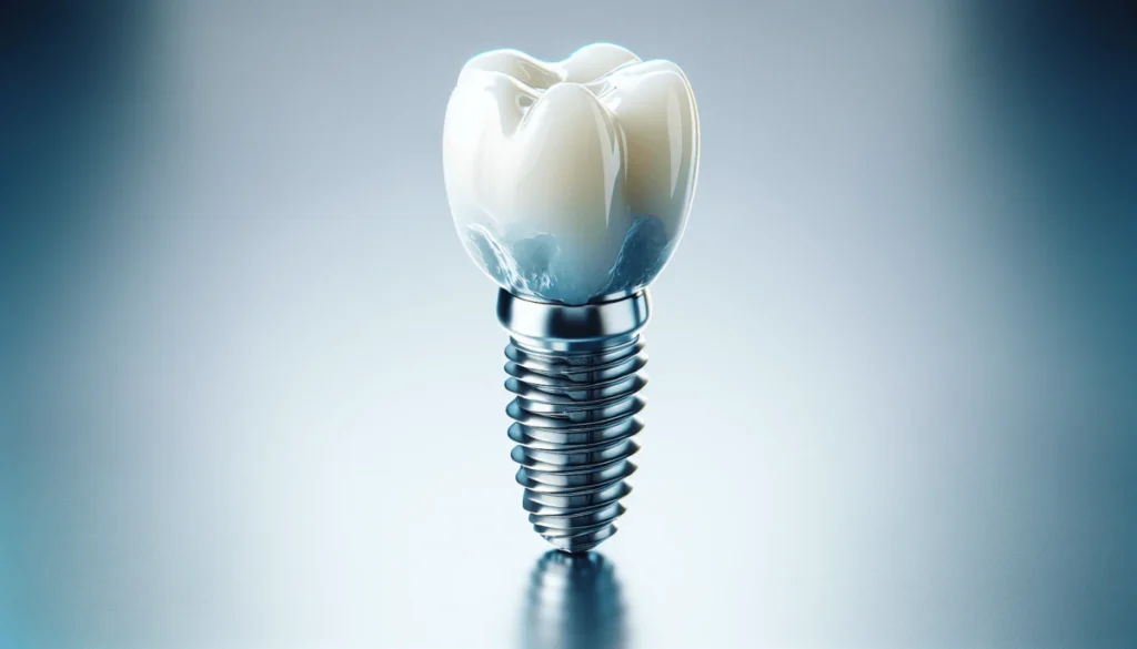
The clinical landscape of restorative dentistry has undergone a fundamental transformation, moving from reactive maintenance to a model of bio-integrated permanence. At the center of this shift is the dental implant, a titanium or zirconia root analog that serves as a structural foundation for oral rehabilitation. While the surgical hardware is often the focus of patient interest, the true determinant of long-term success is the underlying architectural strategy. This involves a synthesis of osseointegration physics, prosthetic design, and financial engineering, all of which must be aligned to ensure the restoration remains viable for the duration of a human lifespan.
Navigating the contemporary market for oral reconstruction requires an analytical understanding of how “plans” are structured. In this context, a plan is not merely a financial instrument or an insurance policy; it is a comprehensive clinical roadmap that accounts for bone density, systemic health variables, and the mechanical loads of mastication. As we move through 2026, the complexity of these frameworks has increased, driven by advancements in guided surgery, 3D bone grafting, and the integration of digital occlusal analysis. The divergence between a standardized, volume-based approach and a precision-engineered clinical strategy has never been more pronounced.
For the serious patient or the medical professional, identifying the optimal path forward requires moving past surface-level marketing. One must analyze the structural integrity of the proposed solution, the biological capacity of the recipient site, and the long-term adaptability of the prosthetic components. This editorial reference provides a definitive exploration of the modern restorative landscape, prioritizing technical nuance and clinical honesty over the simplified narratives often found in the consumer dental space.
Understanding “top dental implant plans.”
To engage with the concept of top dental implant plans, one must first decouple the idea of “planning” from simple insurance coverage. In a professional dental architecture context, a premier plan is a functional ecosystem. It represents a geographic and clinical location that supports the surgical integrity of the implant while providing the logistical infrastructure required for lifelong maintenance. A plan may be aesthetically brilliant in its initial presentation, but if it lacks a roadmap for crestal bone preservation or prosthetic repair, it fails to meet the criteria for a high-tier restorative strategy.
From a clinical perspective, these plans are evaluated based on their “Biological Margin,” the amount of bone and soft tissue preserved during and after the surgical phase. From a logistical perspective, they are judged by the interoperability of their components; for instance, using “open-platform” components that allow for future repairs by any qualified clinician globally, rather than proprietary systems that lock the patient into a single provider network.
Oversimplification Risks
The most significant risk in dental planning is the “Immediate Gratification Trap.” An oversimplified view often suggests that “Teeth in a Day” or immediate loading is the standard of care for every patient. This ignores the biological necessity of primary stability and the potential for micro-motions to disrupt osseointegration in patients with compromised bone density. A professional assessment prioritizes “Biological Resilience,” choosing plans that offer staged approaches when the recipient site lacks the structural volume to support immediate mechanical loads.
Contextual Background: The Evolution of Bio-Integrated Restorations
The history of dental implants is rooted in the mid-20th-century discovery of osseointegration by Per-Ingvar Brånemark, who observed that titanium could fuse with bone without causing a foreign-body rejection. This era established the “Heritage” standard: external hex connections and machined surfaces. These early plans were largely focused on survival—simply ensuring the metal stayed in the jaw.
By the early 2000s, the focus shifted toward “Platform Switching” and surface roughening (SLA or RBM), which encouraged faster bone growth and better soft-tissue attachment. Following this, the development of Cone Beam Computed Tomography (CBCT) revolutionized the planning phase, moving from two-dimensional X-rays to three-dimensional surgical guides. Today, in 2026, the evolution is driven by materials science,e where zirconia implants are gaining traction for patients seeking metal-free alternatives, and digital workflows allow for the “Reverse Planning” of restorations, where the final tooth position dictates the implant placement rather than the other way around.
Conceptual Frameworks and Mental Models for Evaluation
Experienced prosthodontists utilize specific mental models to evaluate the viability of a restorative plan before the first incision is made.
1. The Force-to-Surface-Area (FSA) Model
This model evaluates a plan based on the mechanical load the implant will bear. In the posterior mandible, where chewing forces are highest, the plan must prioritize wider implant diameters or “Double-Implant” configurations for single large molars. In the anterior maxilla, where forces are lower but aesthetic demands are higher, the FSA model shifts to prioritize soft-tissue volume and “Emergence Profile.”
2. The Bone-Volume-to-Implant-Ratio (BVI)
This posits that the utility of an implant is directly proportional to the amount of healthy bone surrounding it. If a site lacks at least 1.5mm of buccal bone, the BVI framework suggests a “Bone-First” approach (grafting) rather than a “Compromised Placement” approach. The optimal plan aligns the surgical hardware with the biological capacity of the site.
3. The “Serviceability” Framework
As restorations age, parts will wear out. This model favors “Screw-Retained” over “Cement-Retained” prosthetics. Screw-retained plans allow a clinician to easily remove the crown for hygiene or repair without damaging the underlying implant. Cement-retained plans, while often more aesthetic, carry the risk of “Peri-implantitis” caused by excess cement remnants below the gumline.
Key Categories and Variations in Planning
The restorative landscape is categorized into distinct “Operational Profiles,” each with its own trade-offs.
| Profile | Primary Benefit | Significant Constraint | Ideal Candidate |
| Single-Stage Guided | Minimally invasive; fast recovery. | Requires high initial bone volume. | Healthy jaw; single tooth loss. |
| Full-Arch (Fixed) | Immediate function; total transformation. | High mechanical stress on 4-6 posts. | Edentulous (no teeth) patients. |
| Zirconia (Ceramic) | High aesthetics; hypoallergenic. | Lower historical data on fatigue. | Thin tissue; metal sensitivity. |
| Overdenture (Snap-on) | Cost-effective; easier hygiene. | Not fixed; requires removal at night. | Moderate bone loss; budget-conscious. |
| Staged Grafting | Maximum long-term stability. | Long duration (6-12 months). | Significant atrophy; smokers. |
Realistic Decision Logic
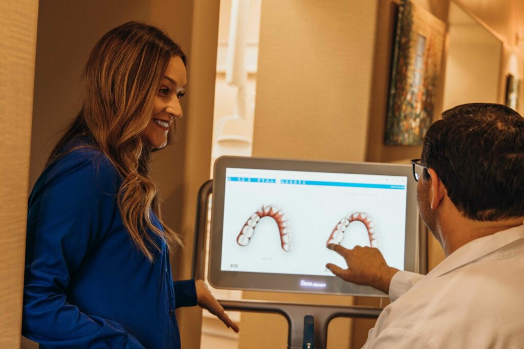
The selection of a profile should be driven by the Patient’s Systemic Health. A patient with uncontrolled diabetes or a history of heavy smoking should avoid “Immediate Loading” profiles, which have a significantly higher failure rate in compromised healing environments. Conversely, a healthy non-smoker with ample bone is an ideal candidate for “Digital Guided Surgery,” which utilizes 3D-printed templates to place implants with sub-millimeter precision.
Detailed Real-World Scenarios and Decision Logic
The Posterior Single-Molar Replacement
A patient seeks to replace a missing lower molar.
-
Decision Point: Should the clinician use a standard 4mm implant or a wide 5mm+ platform?
-
Analysis: The FSA model dictates that the wider platform is necessary to manage the 200+ lbs of force generated during bruxism (grinding).
-
Failure Mode: Under-sizing the implant, leading to “Fatigue Fracture” of the metal post or bone loss due to excessive stress (overloading).
The Anterior “Aesthetic Zone” Reconstruction
A patient has lost a front tooth due to trauma.
-
Constraint: Thin “Biotype” (thin gum,s), which increases the risk of the gray titanium showing through.
-
Decision Point: Selecting a zirconia abutment or a full zirconia implant.
-
Second-Order Effect: The need for a “Connective Tissue Graft” (CTG) to thicken the gums, which adds cost and healing time but ensures the restoration remains invisible at the gumline for decades.
Planning, Cost, and Resource Dynamics
The financial dynamics of restorative dentistry are influenced by regional laboratory costs and the “Biological Overhead” of grafting.
Range-Based Operational Cost Table (US Estimates 2026)
| Phase | Standard Fee Range | Premium/Specialist Fee | Variability Factors |
| Diagnostics (CBCT/Scans) | $300 – $600 | $800 – $1,200 | Software sophistication. |
| Surgical Placement | $1,800 – $3,000 | $3,500 – $5,500 | Guided vs. Freehand. |
| Bone/Sinus Grafting | $600 – $2,500 | $3,000 – $6,000 | Material source (Bio-Oss). |
| Custom Abutment/Crown | $1,200 – $2,500 | $3,000 – $4,500 | Laboratory (Digital vs. Hand). |
Note: Opportunity cost is a significant factor. Choosing a “Budget” plan that lacks a surgical guide may save $500 initially, but results in a “Misaligned Angle” that makes cleaning difficult, leading to a $5,000 failure and replacement cost five years later.
Support Systems, Tools, and Strategic Resources
A successful restoration relies on an ecosystem of specialized support:
-
CBCT Imaging: The “Eyes” of the clinician; essential for avoiding nerve canals and sinus cavities.
-
Intraoral Scanners: Replacing “Goopy” impressions with high-precision 3D digital maps for better crown fit.
-
3D Surgical Guides: Templates that lock the drill into a pre-planned trajectory, eliminating human error during placement.
-
Photogrammetry: Specifically for full-arch cases, ensuring the final bridge fits perfectly across multiple implants.
-
Water Flossers & Interdental Tools: The “Logistical Tether” for the patient to prevent peri-implant disease.
-
Bio-Active Materials: Using Platelet-Rich Fibrin (PRF) drawn from the patient’s own blood to accelerate bone healing.
Risk Landscape and Failure Modes
Even prestigious plans harbor compounding risks.
-
Peri-implantitis: The dental version of gum disease. Unlike natural teeth, implants lack a periodontal ligament (PDL) and its accompanying blood supply, making infection spread much faster toward the bone.
-
Mechanical Fatigue: The screw that holds the crown to the implant can loosen over time. If not tightened promptly, the resulting vibration can fracture the internal threads of the implant.
-
Systemic Changes: A patient who starts a plan as a healthy individual but develops osteoporosis or starts taking bisphosphonates (bone density medication) may face “Late Failure” modes.
Governance, Maintenance, and Long-Term Adaptation
For those with permanent restorations, a “Review Cycle” is necessary.
-
Annual Radiographic Audit: Checking for the first 0.5mm of bone loss, which is the “Leading Indicator” of potential failure.
-
Occlusal Calibration: As natural teeth wear down or shift, the implant (which is fused to bone and cannot move) may begin to take “Heavy Hits” during chewing. Annual adjustment of the bite is critical.
-
Adjustment Triggers: If a patient notices a “Click” or a “Shadow” at the gumline, the plan must pivot to an immediate clinical assessment to prevent the loss of the entire fixture.
Measurement, Tracking, and Evaluation Signals
How do you measure the success of a dental plan?
-
Leading Indicators: High “Insertion Torque” (35Ncm+); zero bleeding on probing; stable bone levels at the one-year mark.
-
Qualitative Signals: Patient comfort during mastication of tough foods; lack of “Metallic Taste” (which can signal galvanic corrosion or infection).
-
Documentation: Requesting the “Implant Passport”—a document containing the exact serial number, size, and brand of the hardware used, ensuring future serviceability by any doctor.
Common Misconceptions and Oversimplifications
-
“Implants are Forever”: They are “Permanent” only if the biological environment is maintained. Without hygiene, they can fail faster than natural teeth.
-
“Generalists are the Same as Specialists”: While many general dentists place implants, specialists (Periodontists/Oral Surgeons) have 3-4 years of additional residency focused exclusively on bone and soft tissue.
-
“Titanium is Always Safe”: While rare, some patients exhibit “Titanium Sensitivity,” manifesting as unexplained inflammation; zirconia is the 2026 hedge for these cases.
-
“The Cheapest Brand is Fine”: “Generic” implant companies often go out of business, leaving patients with “Orphan Implants” for which no repair parts exist.
-
“Smoking is a Minor Risk”: Nicotine constricts blood flow; smoking increases the risk of implant failure by nearly 300%.
-
“Pain is the First Sign of Failure”: Because implants lack nerves, bone loss is usually painless. By the time it hurts, the implant is often already loose.
Conclusion
The architecture of a permanent smile is a strategic exercise in aligning medical engineering with human biology. It is a transition from reactive “Patchwork” dentistry to a proactive, bio-integrated system. Whether one is replacing a single tooth or undergoing a full-mouth reconstruction, success depends on the alignment of technical preparation, material quality, and lifelong maintenance. In 2026, the ultimate luxury in dentistry is not the crown itself, but the predictability of the underlying process, and the confidence that the restoration will remain a functional part of the body’s anatomy for decades to come.