Diagnostic Imaging Plans: The Definitive 2026 Editorial Reference
The modern clinical landscape is increasingly defined not by the intervention itself, but by the quality of the visual intelligence preceding it. Diagnostic imaging has moved beyond the peripheral role of “confirming a hunch” to become the central nervous system of medical decision-making. In 2026, the resolution of an image, whether captured through magnetic resonance, positron emission, or computed tomography, serves as the definitive map for surgical precision, oncological monitoring, and preventive longevity. However, the sheer volume of available modalities has created a paradox of choice; the challenge today is not merely gaining access to technology, but the strategic application of that technology to a specific biological inquiry.
Effective clinical governance requires a transition from “ordering a scan” to architecting a visual roadmap. A sophisticated diagnostic strategy must account for the intricate interplay between spatial resolution, contrast sensitivity, and the inherent biological “noise” of the human body. As we move deeper into an era of personalized medicine, the distinction between routine screening and high-resolution diagnostic investigation has never been more critical. The stakes are particularly high in the detection of early-stage pathologies, where a slight variation in the sequence or the strength of a magnetic field can determine whether an abnormality is addressed or overlooked.
Understanding “diagnostic imaging plans.”

To effectively engage with diagnostic imaging plans, one must first decouple the procedure from the objective. In a professional radiological context, a plan is a longitudinal protocol designed to answer a specific clinical hypothesis. It is not an isolated event but a structured journey that may include baseline scans, interval monitoring, and post-intervention verification. A plan might utilize the world’s most powerful 7-Tesla MRI, but if it lacks clinical contextualization, in the correlation between pixels on a screen and a patient’s physiological symptoms, ms it fails the criteria of a high-tier diagnostic strategy.
Multi-Perspective Explanation
From a physics perspective, these plans are judged by their signal-to-noise ratio (SNR). The objective is to maximize the clarity of the target tissue while minimizing artifacts from movement or blood flow. From a biomechanical perspective, evaluation is based on temporal resolution, in the ability to capture motion, such as the beating of a heart or the expansion of a lung. Finally, from a regulatory perspective, a plan must be scrutinized for its adherence to the ALARA principle (As Low As Reasonably Achievable), ensuring that the diagnostic benefit of ionizing radiation always outweighs the biological cost of exposure.
Oversimplification Risks
The primary risk in diagnostic planning is modality reductionism—the belief that all scans within a category are interchangeable. In reality, there are hundreds of different sequences (T1, T2, FLAIR, DWI) that can be applied to a single MRI session. An oversimplified view often ignores the importance of coil selection or contrast kinetics. A professional assessment avoids these pitfalls by prioritizing protocol optimization, ensuring the scanner is tuned specifically to the tissue being examined, rather than relying on factory-default settings.
Contextual Background: The Evolution of Medical Visualization
The history of medical imaging has moved from the “Shadow Era”—beginning with Wilhelm Röntgen’s discovery of X-rays in 1895—to the cross-sectional era of the 1970s, and now into the molecular and functional era of 2026. The invention of Computed Tomography (CT) by Godfrey Hounsfield allowed clinicians to see inside the body in three dimensions for the first time, effectively “slicing” the patient without a scalpel.
By the 1980s, Magnetic Resonance Imaging (MRI) introduced the ability to visualize soft tissues with incredible contrast without using ionizing radiation. Today, the evolution is driven by radiomics and hybrid imaging, such as PET/MRI.
Conceptual Frameworks and Mental Models for Evaluation
Strategic radiologists and clinicians utilize specific frameworks to evaluate the viability of an imaging roadmap.
1. The Anatomical vs. Functional Framework
This model posits that every imaging question is either about structure (is the bone broken?) or process (is the blood flowing?). A top-tier plan often combines both. For example, a structural MRI of the heart tells you about the size of the chambers, but a functional cardiac MRI with stress testing tells you if the muscle is actually receiving enough blood under load.
2. The Window of Resolution Mental Model
In this model, every imaging tool has a lowest limit of what it can perceive. A CT scan might miss a microscopic tumor that a PET scan catches, while an ultrasound might see a gallstone that an MRI misses due to the specific physics of calcium visualization. The best plans choose the tool based on the expected size and density of the pathology.
3. The Cost-Benefit Hemodynamics Logic
This framework evaluates the invasiveness of the plan. A non-invasive CT Angiogram is safer and faster for initial screening, but an invasive catheter angiogram allows for immediate intervention, such as placing a stent. This model determines if the imaging should be an observational or a therapeutic event.
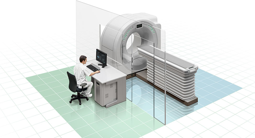
Key Categories: Modalities and Mechanical Trade-offs
The diagnostic landscape is categorized into distinct operational profiles based on the energy used to create the image.
| Modality | Energy Source | Primary Benefit | Significant Constraint |
| CT Scan | Ionizing Radiation | Incredible speed; best for bone/lung. | High radiation; poor soft tissue contrast. |
| MRI | Magnetic Fields | Superior soft tissue; no radiation. | Slow; loud; no ferrous metal allowed. |
| Ultrasound | Sound Waves | Real-time, portable, and safest. | Operator dependent; blocked by bone/air. |
| PET Scan | Radioactive Tracers | Shows metabolic activity/cancer. | Low anatomical detail; very expensive. |
| X-Ray | Ionizing Radiation | Cheap; fast; guided injections. | 2D only; limited diagnostic depth. |
| Nuclear Med | Gamma Rays | Organ function (Kidney/Thyroid). | Requires injection of isotopes. |
Realistic Decision Logic
The selection of a profile must be driven by the target tissue density. If the question involves the hardware of the body (bones, joints, lungs), a CT-based plan is often superior. If the question involves the software (brain, nerves, ligaments, organs), an MRI-based plan is usually mandatory. For emergency scenarios where every second counts—such as a suspected stroke or internal bleeding—the speed of a CT scan outweighs the higher resolution of an MRI.
Detailed Real-World Scenarios and Decision Logic
The Chronic Neurological Search
A 35-year-old with unexplained tingling and vision loss.
-
Decision Point: CT Head vs. MRI Brain with Contrast.
-
Analysis: A CT will likely come back “normal” even if the patient has active Multiple Sclerosis.
-
Outcome: The plan utilizes a 3-Tesla MRI with GAD Contrast, looking specifically for white matter lesions that are invisible to X-ray-based technology.
High-Stakes Cardiac Clearance
A 55-year-old executive with high cholesterol wants to know their actual heart attack risk.
-
Constraint: Asymptomatic, but high familial risk.
-
Decision Point: Stress Test (ECG) vs. Coronary CT Angiography (CCTA).
-
Second-Order Effect: A stress test primarily finds blockages over 70%. A CCTA with AI plaque analysis can see 10% blockages and identify “soft plaque” prone to rupture, allowing for a primary prevention strategy.
Planning, Cost, and Resource Dynamics
The financial dynamics of diagnostic imaging are defined by hardware amortization and interpretive expertise.
Range-Based Operational Cost Table (US Estimates 2026)
| Procedure | Standard Facility Fee | High-Resolution Tier | Key Cost Driver |
| X-Ray (Standard) | $100 – $300 | $400 – $800 | Digital vs. Analog. |
| Ultrasound | $300 – $800 | $1,000 – $2,500 | 3D/4D Rendering. |
| CT Scan (Standard) | $800 – $2,500 | $3,000 – $6,000 | Contrast & AI Analysis. |
| MRI (1.5T or 3T) | $1,500 – $4,500 | $5,000 – $12,000 | Sequencing time; sedation. |
| PET/CT | $4,000 – $7,000 | $8,000 – $15,000 | Isotope half-life/cost. |
In 2026, the best value is often found in independent imaging centers rather than hospital-based systems. Hospital fees often include facility charges that can double or triple the cost of an identical scan performed at a standalone clinic.
Support Systems, Tools, and Strategic Resources
A successful visualization program relies on a digital stack of specialized resources:
-
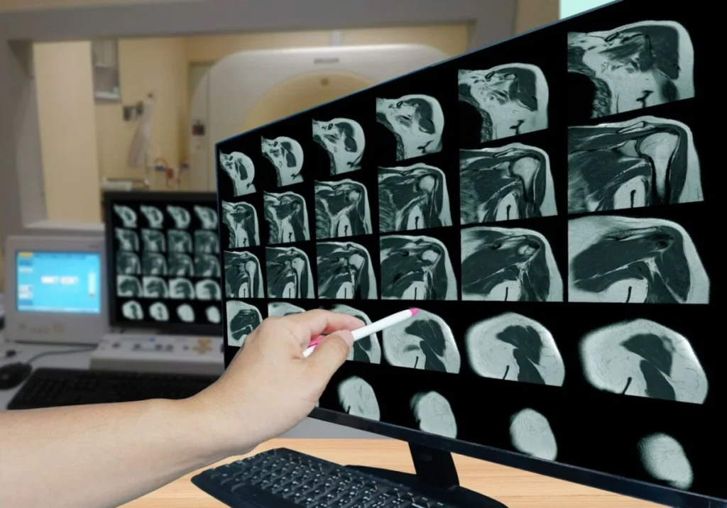
PACS (Picture Archiving and Communication System): The cloud infrastructure where high-resolution images are stored and shared between specialists.
-
AI-Assisted Triage: Software that scans images for life-threats, such as a brain bleed, and moves them to the top of the radiologist’s queue.
-
3D Reconstruction Labs: Converting flat CT slices into three-dimensional models for surgical planning.
-
Radiomics Engines: Tools that extract thousands of data points from an image that the human eye cannot perceive, such as texture analysis of a tumor.
-
Dose-Tracking Software: A governance tool that monitors a patient’s lifetime radiation exposure to prevent cumulative risk.
-
Contrast Power-Injectors: Precise machines that time the injection of dye to the exact millisecond the heart pumps it into the target organ.
Risk Landscape and Failure Modes
Even the most prestigious diagnostic imaging plans harbor compounding risks.
-
The Incidentaloma Trap: Finding a harmless spot on an organ that leads to unnecessary surgery or years of stressful monitoring.
-
Contrast-Induced Nephropathy: The risk that the dye used in scans can damage the kidneys in patients with underlying renal disease.
-
Motion Artifacts: If a patient moves during a 45-minute MRI, the entire plan may be unreadable, leading to a non-diagnostic report.
-
Magnetic Projectiles: In the MRI suite, any ferrous object becomes a lethal missile, representing a significant safety failure mode.
Governance, Maintenance, and Long-Term Adaptation
To maintain the diagnostic utility of a plan, the medical team must adopt a governance mindset.
-
The Interval Integrity Audit: For chronic conditions, such as a small lung nodule, the timing of the next scan is more important than the scan itself. A plan must adhere to strict guidelines to avoid over-scanning.
-
Review Cycles: Every 12-24 months, the imaging protocol should be reviewed to see if newer, safer technologies, such as moving from CT to MRI, have become available.
-
Checklist for Accuracy:
Measurement, Tracking, and Evaluation Signals
How do you measure the success of a diagnostic imaging plan?
-
Leading Indicators: Time-to-report (how fast the clinician gets the answer); image quality score (absence of blur or noise).
-
Qualitative Signals: Diagnostic confidence—the ability of a surgeon to say, “I know exactly where the lesion is and what it is touching.”
-
Documentation Examples: A radiological passport that includes the digital access to all raw DICOM files, the formal report, and the specific technique used, such as 1.5mm slices with intravenous contrast.
Common Misconceptions and Oversimplifications
-
Radiation Stays in the Body: False. X-rays pass through you like light through glass. Once the machine is off, the radiation is gone.
-
A Normal Scan Means Total Health: A scan only shows what it is designed to see. A normal brain MRI doesn’t rule out chemical depression or early-stage Alzheimer’s.
-
MRI is Always Better Than CT: CT is far superior for finding a broken bone or a kidney stone due to the density of the material.
-
I need an MRI for My Back Pain: 90% of back pain resolves without imaging. Imaging too early often leads to unnecessary surgeries for “normal” age-related wear.
-
Contrast is Just Water: Contrast is a pharmaceutical drug with its own risks and side effects. It should only be used when necessary.
-
The Radiologist is Just a Technician: Radiologists are medical doctors who have spent 5+ years specializing only in the interpretation of images. They are the “doctor’s doctor.”
Ethical and Practical Considerations
The ethics of diagnostic imaging in 2026 revolve around data ownership and access equity. Who owns the rights to your 3D heart model—you or the hospital? Furthermore, there is a growing divide between patients who receive AI-enhanced, high-resolution care and those in rural areas limited to 20-year-old hardware. Intellectual honesty requires acknowledging that a diagnostic plan is only as good as the specialist interpreting it; a high-end scan read by an overworked, non-specialized generalist represents a significant clinical risk.
Conclusion
The architecture of medical visualization is a strategic exercise in balancing the search for truth with the preservation of biological safety. It is a transition from guesswork to precision. Whether you are monitoring a life-threatening malignancy or seeking the source of chronic pain, success depends on the integration of physics, clinical judgment, and patient discipline.